Microscope Endodontics
Dental Microscopes provide magnification unavailable to the naked eye. The fiber-optic illumination allows endodontists to see much better than with a regular light. They are also helpful in aiding the endodontists to see the minute details inside your tooth. A camera is attached to the operating microscope allowing us to record images of your tooth to further document the endodontists' findings.
- The improved accuracy offered by the increased magnification and illumination of a microscope allows the endodontists more precision for both cleaning canals and effectively treating endodontic conditions.
- More precision results in reduced trauma to the mouth during a procedure, as well as less discomfort during and after the procedure. Reduced trauma translates to easier healing and a reduced chance of needing additional treatment.
- Increased magnification allows Dr. Langella to use smaller instruments. By using smaller instruments, she can make more precise incisions and sutures. This minimizes the surgical site, which in turn minimizes discomfort during and after the procedure.
- As Dr. Langella uses the microscope to see better, she can more easily detect, diagnose, treat and prevent more serious conditions.
- The heightened efficiency saves you money on potential extra or more serious treatments, treatments that Dr. Langella can avoid as a result of this technology.
Cone Beam CT Scanner
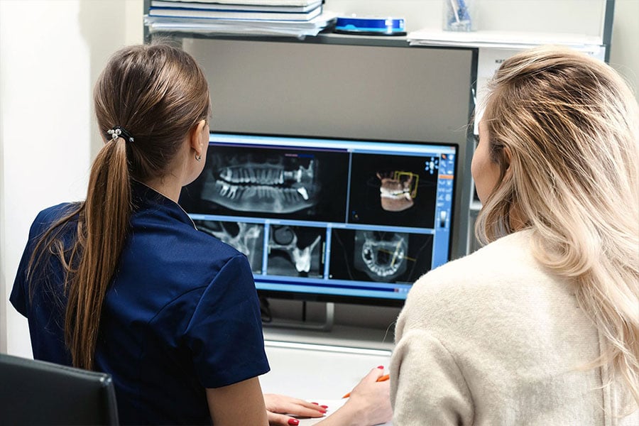
Our virtually painless dental office is one of the few dental offices with an on-site 3D Cone Beam CT Scanner! This revolutionary technology provides three-dimensional views allowing Dr. Langella to thoroughly assess and plan each patient before any procedure. Unlike a traditional CT Scanner, this 3D system utilizes Cone Beam Technology and provides us with precise, crystal-clear images. The information the scan offers is invaluable during the assessment and planning phases of root canals and many other surgeries. We can accurately visualize and prepare for issues such as bone quality and the location of sinuses, nerves, and other vital anatomical landmarks. Utilizing this scan before surgery greatly improves our patients’ experience and delivers a positive treatment outcome.
Digital Radiography
Digital x-rays are the newest technology used in dentistry to take and archive dental x-rays. Digital x-rays significantly reduce the amount of radiation as compared to traditional dental x-rays. This technique captures a digital picture of teeth with their supporting bone structures and stores the images on a computer in our dental office. You and your endodontist will be able to instantly view your x-rays and enlarge the image to aid in the identification of dental problems and to gauge your dental health. Your endodontist will use this information to create an individualized treatment plan.
Intra-Oral Camera
Taking pictures of the teeth and supporting tissue with a small video camera about the size of a pen is a wonderful addition to dentistry today. The intra-oral camera creates digital images that can be stored on a computer. The images are shared with our patients so they can join us in the "co-diagnosis" of problems with their dental health. Your endodontist can show you how others view your smile and which dental fillings are broken or discolored. We look forward to showing you a dental tour of your mouth in the privacy of your treatment room.